|
Overview
 The common bunion is a localized area of enlargement of the inner portion of the joint at the base of the big toe. The enlargement actually represents a misalignment of the big toe joint (metatarsal phalangeal joint) and, in some cases, additional bone formation. The misalignment causes the big toe to point outward (medically termed hallux valgus deformity) toward the smaller toes. This deformity is progressive and will increase with time. The enlarged joint at the base of the big toe (the first metatarsophalangeal joint, or MTP joint) can become inflamed with redness, tenderness, and pain. A small fluid-filled sac (bursa) adjacent to the joint can also become inflamed (bursitis), leading to additional swelling, redness, and pain. A more deep joint pain may occur as localized arthritis develops in later stages of the deformity. A less common bunion is located at the joint at the base of the smallest (fifth) toe. This bunion is sometimes referred to as a tailor's bunion or bunionette. The common bunion is a localized area of enlargement of the inner portion of the joint at the base of the big toe. The enlargement actually represents a misalignment of the big toe joint (metatarsal phalangeal joint) and, in some cases, additional bone formation. The misalignment causes the big toe to point outward (medically termed hallux valgus deformity) toward the smaller toes. This deformity is progressive and will increase with time. The enlarged joint at the base of the big toe (the first metatarsophalangeal joint, or MTP joint) can become inflamed with redness, tenderness, and pain. A small fluid-filled sac (bursa) adjacent to the joint can also become inflamed (bursitis), leading to additional swelling, redness, and pain. A more deep joint pain may occur as localized arthritis develops in later stages of the deformity. A less common bunion is located at the joint at the base of the smallest (fifth) toe. This bunion is sometimes referred to as a tailor's bunion or bunionette.Causes Bunions form when the normal balance of forces exerted on the joints and tendons of your feet are disrupted. This can lead to instability in the big toe joint - also known as the first metatarsophalangeal (MTP) joint, causing a deformity. Bunions develop over years of abnormal motion and pressure on your big toe joint. They often result from a combination of your inherited foot type, faulty foot mechanics that affect the way you walk and shoes that fit improperly. Other causes of bunions include foot injuries. Deformities present at birth (congenital). Neuromuscular disorders, such as cerebral palsy or post- polio syndrome (post-poliomyelitis). Bunions may be associated with various forms of arthritis, including inflammatory or degenerative, causing the protective cartilage that covers your big toe joint to deteriorate. An occupation that puts extra stress on your feet also can be a cause. Waiters, factory workers, dancers and athletes often are more prone to developing bunions. Symptoms Symptoms include redness, swelling and pain which may be present along the inside margin of the foot. The patients feet may become too wide to fit into their normal size shoes and moderate to severe discomfort may occur when the patient is wearing tight shoes. A "hammer toe" may occur at the 2nd toe. This is when the toe contracts and presses on the shoe. Subsequently, this may cause a corn on top of the 2nd toe. Diagnosis When an x-ray of a bunion is taken, there is usually angulation between the first metatarsal bone and the bones of the big toe. There may also be angulation between the first and second metatarsal bones. These angular irregularities are the essence of most bunions. In general, surgery for bunions aims to correct such angular deformities. Non Surgical Treatment Wearing the right shoes, using shoe inserts (orthoses) and padding, and taking painkillers can all help to ease your symptoms of a bunion. However, these treatments can?t cure a bunion or stop it getting worse. If you have severe pain or discomfort from a bunion, you may be advised to have an operation to correct it. One of the most important things you can do is to wear the right footwear. You should try to wear flat, wide-fitting shoes with laces or an adjustable strap that fits you properly. You may also want to place a bunion pad over your bunion to give it some protection from the pressure of your shoes. You can usually buy these pads from a pharmacy, or get them from your podiatrist or chiropodist. He or she may also recommend a shoe insert, which can be moulded specifically to your foot. Shoe inserts aim to reduce the pain of your bunion by improving how you walk. You can take over-the-counter painkillers, such as paracetamol or ibuprofen, to help relieve the pain and inflammation of your bunion. Always follow the instructions in the patient information leaflet that comes with your medicine. Medicines give temporary relief but they won?t be able to cure your bunion or prevent it from getting worse. If you have a bunion as a result of underlying arthritis, your doctor may prescribe specific medicines to treat this. 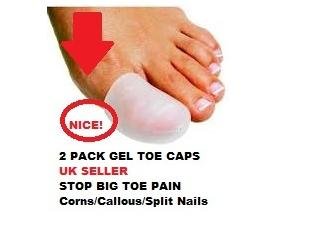 Surgical Treatment Bunion surgery is usually done as an out patient procedure, so the patient does not have to stay in hospital overnight although it is usually performed under a general anesthetic. The procedure involves the surgeon making a cut on the inside of the big toe joint and removing excess bone whilst also repositioning ligaments and tendons. The joint may be fixed with screws or wires, which may be dissolve, or may be removed at a later date or in some cases, remain in the foot permanently. After the operation the foot will be immobilized, often in a cast for 4 to 8 weeks to keep the bones in alignment. Crutches will usually be issued to help the patient get around. After this period, the foot will be assessed to check the bones have healed correctly. At which point full weight bearing may be gradually introduced. Overview
Pronation is the natural motion of the foot as it roles inward after the foot makes contact with the ground. It gives the foot the opportunity to act as a shock absorber for the body and adapt to the contour of the ground. Too much pronation will cause the arch of the foot to flatten excessively placing stress and pressure on tissues and ligaments of the foot. Over pronation can lead to numerous foot problems including plantar fasciitis, ankle pain, shin pain, knee pain, bunions, mortons neuroma, chondromalcia patallae and lower back pain.  Causes A common cause of pronation is heredity - we can inherit this biomechanical defect. The second most common cause is due to the way our feet were positioned in the uterus while we were developing; this is called a congenital defect. In either instance, the following occurs in our feet during our development. Symptoms Common conditions that develop with prolonged overpronation typically include plantar fasciitis, achilles tendonitis, shin splints, posterior tibial stress syndrome and even IT band syndrome. With long term neglect you may see the development of bunyons, foot deformities and early onset of hip and knee arthritis. Diagnosis You can test for pronation by looking at the leg and foot from the back. Normally you can see the Achilles Tendon run straight down the leg into the heel. If the foot is pronated, the tendon will run straight down the leg, but when it lies on the heel it will twist outward. This makes the inner ankle bone much more prominent than the outer ankle bone.  Non Surgical Treatment Overpronation of the feet can be corrected in some cases and in others it can be effectively managed. Overpronators can train themselves to change their running gait, wear arch supports, orthotic insoles or specialist shoes for overpronators. In order to determine exactly what is happening during the stride, it is necessary to have a gait analysis conducted by a professional. The extent of overpronation can then be determined, and the causes can be identified and corrected directly. The main corrective methods used for excessive pronation are orthotics. Orthotics are the most straightforward and simplest solution to overpronation. Orthotics are devices which can be slipped into shoes which will offer varying degrees of correction to the motion of the foot. Orthotics help to support the arches and distribute the body weight effectively, and are usually the best treatment choice for moderate to severe overpronation. Orthotics may require existing insoles to be removed from your shoes to accommodate them; although most running shoes will have a removable insole to accommodate an orthotic insole. Prevention Custom-made orthotics supports not only the arch as a whole, but also each individual bone and joint that forms the arch. It is not enough to use an over-the-counter arch support, as these generic devices will not provide the proper support to each specific structure of the arch and foot. Each pronated foot?s arch collapses differently and to different degrees. The only way to provide the support that you may need is with a custom-made device. This action of the custom-made orthotic will help to prevent heel spurs, plantar fasciitis, calluses, arch pain, and weakness of the entire foot. Overview
Sever disease, first described in 1912, is a painful inflammation of the calcaneal apophysis. It is classified with the child and adolescent nonarticular osteochondroses. (The other disease in this group is Iselin disease, which is inflammation of the base of the fifth metatarsal.) The etiology of pain in Sever disease is believed to be repetitive trauma to the weaker structure of the apophysis, induced by the pull of the tendo calcaneus (Achilles tendon) on its insertion. This results in a clinical picture of heel pain in a growing active child, which worsens with activity. Sever disease is a self-limited condition, accordingly, no known complication exists from failure to make the correct diagnosis. Causes The usual cause is directly related to overuse of the bone and tendons in the heel. This can come from playing sports or anything that involves a lot of heel movement. It can be associated with starting a new sport, or the start of a new season, or too much weight bearing on the heel. Also, excessive traction could cause this, since the bones and tendons are still developing. Many children who over pronate their feet exhibit symptoms and in most patients, it usually involves both heels. Symptoms This syndrome can occur unilaterally or bilaterally. The incidence of bilaterally is approximately 60%. Common signs and symptoms include posterior inferior heel pain (over the medial and lateral surface of the bone). Pain is usually absent when the child gets up in the morning. Increased pain with weight bearing, running or jumping (= activity-related pain). The area often feels stiff. The child may limp at the end of physical activity. Tenderness at the insertion of the tendons (= an avascular necrosis of the arthropathy). Limited ankle dorsiflexion range secondary to tightness of the Achilles tendon. Hard surfaces and poor-quality or worn-out athletic shoes contribute to increased symptoms. The pain gradually resolves with rest. Reliability or validity of methods used to obtain the ankle joint dorsiflexion or biomechanical malalignment data are not commented upon, thus reducing the quality of the data. Although pain and limping are mentioned as symptomatic traits, there have been no attempts to quantify the pain or its effect on the individual. Diagnosis A physical exam of the heel will show tenderness over the back of the heel but not in the Achilles tendon or plantar fascia. There may be tightness in the calf muscle, which contributes to tension on the heel. The tendons in the heel get stretched more in patients with flat feet. There is greater impact force on the heels of athletes with a high-arched, rigid foot. Non Surgical Treatment Treatment is primarily directed towards reducing the amount of stress to the heel. Often a heel lift, is placed in the shoe to reduce the pull of the Achilles tendon on the apophysis. Gel or cushioned heel cups may also be helpful in reducing micro trauma to the heel. Orthotic control may also be indicated when a pathologic condition exists in the foot that may be contributing to the increased heel stress. Occasionally, it becomes necessary for adequate healing, to rest the area completely. This can be accomplished either by complete elimination of all strenuous activities, or by using a walking cast or crutches. Often simply reducing activity levels is adequate. Your physician will discuss the best treatment plan with you and your child. Overview
Plantar fasciitis refers to an inflammation of the plantar fascia, a thick, fibrous band running along the sole of the foot. Such inflammation results from direct injury to the plantar fascia, usually, repeated trauma to the tissue where the fascia attaches to the calcaneus or heel bone. The plantar fascia is critical in maintaining the foot?s complex arch system, also playing a role in balance and fine control of certain phases of the athlete?s gait. Injury to the plantar fascia is particularly painful and disabling for runners and can often prove stubbornly resistant to treatment. Rehabilitation is frequently a lengthy and frustrating process. For these reasons, care should be taken where possible to avoid such injury by means of preventative exercises and sensitivity to early warning signs. 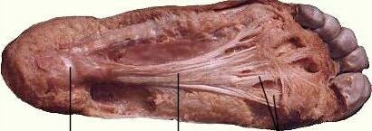 Causes If the feet are exposed to damp conditions for prolonged periods, you can develop Trench Foot. Most commonly associated with World War One, it now tends to be seen in builders, hikers or festival goers. Trench foot causing numbness and bottom of foot pain. With Trench Foot, the sole of the foot turns a white/grey colour and you may develop pins and needles or numbness. Other symptoms include pain in bottom of foot and swelling. If left untreated, you can develop blisters and permanent nerve damage which can lead to the need for amputation. Treatment and prevention aim to reduce the dampness around the foot and ensure good foot hygiene. Slowly rewarming the feet and using special products in a foot bath really helps reduce the damage and foot arch pain from Trench Foot. Symptoms Most flat feet do not cause pain or other problems. Children may have foot pain, ankle pain, or lower leg pain. They should be evaluated by a health care provider if this occurs. Symptoms in adults may include tired or achy feet after long periods of standing or playing sports. Diagnosis Magnetic Resonance Imaging (MRI) can show tendon injury and inflammation but cannot be relied on with 100% accuracy and confidence. The technique and skill of the radiologist in properly positioning the foot with the MRI beam are critical in demonstrating the sometimes obscure findings of tendon injury around the ankle. Magnetic Resonance Imaging (MRI) is expensive and is not necessary in most cases to diagnose posterior tibial tendon injury. Ultrasound has also been used in some cases to diagnose tendon injury, but this test again is usually not required to make the initial diagnosis. Non Surgical Treatment How the pain in the bottom of your foot is treated will depend heavily on the cause of the pain. Diagnosing the pain while it?s in the early stages is important when determining the best treatment options. If the pain is mild to moderate, simple improvements in footwear can help reduce the symptoms. Most patients must use the RICE method for effective treatment. RICE stands for Rest, Ice, Compression, and Elevation. This is a popular treatment used by athletes. It involves resting the foot, icing it for fifteen to twenty minute intervals, compressing the foot with a bandage, and elevating it at least twelve inches above the heart. Ant-inflammatory and pain medications are also sometimes used to treat bottom-of-foot pain. For more serious cases, steroid injections or foot surgery may help reduce pain and swelling and correct the underlying condition (if there is one.) If you suffer from a severe case of plantar fasciitis and non-surgical methods fail, your doctor may recommend cortisone injections to relieve the pain. If cortisone injections fail, your doctor may recommend a surgical procedure that involves cutting and releasing the plantar fascia.  Surgical Treatment Fallen arches may occur with deformities of the foot bones. Tarsal coalition is a congenital condition in which the bones of the foot do not separate from one another during development in the womb. A child with tarsal coalition exhibits a rigid flat foot, which can be painful, notes the patient information website eOrthopod. Surgery may prove necessary to separate the bones. Other foot and ankle conditions that cause fallen arches may also require surgery if noninvasive treatments fail to alleviate pain and restore normal function. Prevention Foot and ankle injuries are common in sports, especially running, tennis and soccer. But sports enthusiasts can decrease the risk of injury by taking some precautions. Lightly stretch or better yet, do a slow jog for two to three minutes to warm up the muscles. Don't force the stretch with a "bouncing motion." The amount of time spent on the activity should be increased gradually over a period of weeks to build both muscle strength and mobility. Cross training by participating in different activities can help build the muscles. People whose feet pronate or who have low arches should choose shoes that provide support in both the front of the shoe and under the arch. The heel and heel counter (back of the shoe) should be very stable. Those with a stiffer foot or high arches should choose shoes with more cushion and a softer platform. Use sport-specific shoes. Cross training shoes are an overall good choice; however, it is best to use shoes designed for the sport. Stretching Exercises Below are two simple plantar fasciitis stretching exercises to help improve the flexibility of the muscles and tendons around the foot and ankle. Plantar fasciitis stretch taken from The Stretching Handbook. Kneel on one foot and place your body weight over your knee. Keep your heel on the ground and lean forward. In the photo to the left, the athlete is stretching the arch of her left foot. Kneel on one foot with your hands on the ground. Place your body weight over your knee and slowly move your knee forward. Keep your toes on the ground and arch your foot. In the photo to the right, the athlete is stretching the arch of his right foot. Overview
Plantar fasciitis refers to an inflammation of the plantar fascia, a thick, fibrous band running along the sole of the foot. Such inflammation results from direct injury to the plantar fascia, usually, repeated trauma to the tissue where the fascia attaches to the calcaneus or heel bone. The plantar fascia is critical in maintaining the foot?s complex arch system, also playing a role in balance and fine control of certain phases of the athlete?s gait. Injury to the plantar fascia is particularly painful and disabling for runners and can often prove stubbornly resistant to treatment. Rehabilitation is frequently a lengthy and frustrating process. For these reasons, care should be taken where possible to avoid such injury by means of preventative exercises and sensitivity to early warning signs. Causes If the feet are exposed to damp conditions for prolonged periods, you can develop Trench Foot. Most commonly associated with World War One, it now tends to be seen in builders, hikers or festival goers. Trench foot causing numbness and bottom of foot pain. With Trench Foot, the sole of the foot turns a white/grey colour and you may develop pins and needles or numbness. Other symptoms include pain in bottom of foot and swelling. If left untreated, you can develop blisters and permanent nerve damage which can lead to the need for amputation. Treatment and prevention aim to reduce the dampness around the foot and ensure good foot hygiene. Slowly rewarming the feet and using special products in a foot bath really helps reduce the damage and foot arch pain from Trench Foot. Symptoms Most flat feet do not cause pain or other problems. Children may have foot pain, ankle pain, or lower leg pain. They should be evaluated by a health care provider if this occurs. Symptoms in adults may include tired or achy feet after long periods of standing or playing sports. Diagnosis Magnetic Resonance Imaging (MRI) can show tendon injury and inflammation but cannot be relied on with 100% accuracy and confidence. The technique and skill of the radiologist in properly positioning the foot with the MRI beam are critical in demonstrating the sometimes obscure findings of tendon injury around the ankle. Magnetic Resonance Imaging (MRI) is expensive and is not necessary in most cases to diagnose posterior tibial tendon injury. Ultrasound has also been used in some cases to diagnose tendon injury, but this test again is usually not required to make the initial diagnosis. Non Surgical Treatment How the pain in the bottom of your foot is treated will depend heavily on the cause of the pain. Diagnosing the pain while it?s in the early stages is important when determining the best treatment options. If the pain is mild to moderate, simple improvements in footwear can help reduce the symptoms. Most patients must use the RICE method for effective treatment. RICE stands for Rest, Ice, Compression, and Elevation. This is a popular treatment used by athletes. It involves resting the foot, icing it for fifteen to twenty minute intervals, compressing the foot with a bandage, and elevating it at least twelve inches above the heart. Ant-inflammatory and pain medications are also sometimes used to treat bottom-of-foot pain. For more serious cases, steroid injections or foot surgery may help reduce pain and swelling and correct the underlying condition (if there is one.) If you suffer from a severe case of plantar fasciitis and non-surgical methods fail, your doctor may recommend cortisone injections to relieve the pain. If cortisone injections fail, your doctor may recommend a surgical procedure that involves cutting and releasing the plantar fascia. Surgical Treatment Fallen arches may occur with deformities of the foot bones. Tarsal coalition is a congenital condition in which the bones of the foot do not separate from one another during development in the womb. A child with tarsal coalition exhibits a rigid flat foot, which can be painful, notes the patient information website eOrthopod. Surgery may prove necessary to separate the bones. Other foot and ankle conditions that cause fallen arches may also require surgery if noninvasive treatments fail to alleviate pain and restore normal function. Prevention Foot and ankle injuries are common in sports, especially running, tennis and soccer. But sports enthusiasts can decrease the risk of injury by taking some precautions. Lightly stretch or better yet, do a slow jog for two to three minutes to warm up the muscles. Don't force the stretch with a "bouncing motion." The amount of time spent on the activity should be increased gradually over a period of weeks to build both muscle strength and mobility. Cross training by participating in different activities can help build the muscles. People whose feet pronate or who have low arches should choose shoes that provide support in both the front of the shoe and under the arch. The heel and heel counter (back of the shoe) should be very stable. Those with a stiffer foot or high arches should choose shoes with more cushion and a softer platform. Use sport-specific shoes. Cross training shoes are an overall good choice; however, it is best to use shoes designed for the sport. Stretching Exercises Below are two simple plantar fasciitis stretching exercises to help improve the flexibility of the muscles and tendons around the foot and ankle. Plantar fasciitis stretch taken from The Stretching Handbook. Kneel on one foot and place your body weight over your knee. Keep your heel on the ground and lean forward. In the photo to the left, the athlete is stretching the arch of her left foot. Kneel on one foot with your hands on the ground. Place your body weight over your knee and slowly move your knee forward. Keep your toes on the ground and arch your foot. In the photo to the right, the athlete is stretching the arch of his right foot.
Overview
 Achilles tendon rupture is most common in people aged 30-50. Patients may describe the injury as feeling or hearing a snap or bang, or as feeling they have been shot in the back of the leg. On examination, patients will have reduced plantarflexion and a positive Thompson test. Surgery is associated with a lower risk of re-rupture and a greater likelihood of returning to sporting activity. Conservative management reduces the chance of complications. Causes The Achilles tendon usually ruptures as a result of a sudden forceful contraction of the calf muscles. Activities such as jumping, lunging, or sprinting can cause undue stress on the Achilles tendon and cause it to rupture. Often there is a background of Achilles tendinitis. Direct trauma to the area, poor flexibility or weakness of the calf muscles or of the Achilles tendon and increasing age are some of the other factors that are associated with an Achilles tendon rupture. Symptoms A sudden and severe pain may be felt at the back of the ankle or calf, often described as "being hit by a rock or shot" or "like someone stepped onto the back of my ankle." The sound of a loud pop or snap may be reported. A gap or depression may be felt and seen in the tendon about 2 inches above the heel bone. Initial pain, swelling, and stiffness may be followed by bruising and weakness. The pain may decrease quickly, and smaller tendons may retain the ability to point the toes. Without the Achilles tendon, though, this would be very difficult. Standing on tiptoe and pushing off when walking will be impossible. A complete tear is more common than a partial tear. Diagnosis Diagnosis is made mostly by clinical examination with a defect usually noted on visual examination and by touching the area. A simple test can be done by squeezing the back of the calf with the foot resting in the air. Normally when squeezing the muscle belly the tendon will shorten causing the foot to move in a downward position. With a rupture this squeezing effect may show no movement of the foot if it is not attached properly. A negative test does not mean there isn't some degree of rupture as some of the tendon fibers may still be attached. Sometimes x-rays, an mri, or an ultrasound can be helpful in determining the extent of the rupture. Non Surgical Treatment Initial treatment for sprains and strains should occur as soon as possible. Remember RICE! Rest the injured part. Pain is the body's signal to not move an injury. Ice the injury. This will limit the swelling and help with the spasm. Compress the injured area. This again, limits the swelling. Be careful not to apply a wrap so tightly that it might act as a tourniquet and cut off the blood supply. Elevate the injured part. This lets gravity help reduce the swelling by allowing fluid and blood to drain downhill to the heart. Over-the-counter pain medication is an option. Acetaminophen (Tylenol) is helpful for pain, but ibuprofen (Motrin, Advil, Nuprin) might be better, because these medications relieve both pain and inflammation. Remember to follow the guidelines on the bottle for appropriate amounts of medicine, especially for children and teens. Achilles tendon rupture is most common in people aged 30-50. Patients may describe the injury as feeling or hearing a snap or bang, or as feeling they have been shot in the back of the leg. On examination, patients will have reduced plantarflexion and a positive Thompson test. Surgery is associated with a lower risk of re-rupture and a greater likelihood of returning to sporting activity. Conservative management reduces the chance of complications. Causes The Achilles tendon usually ruptures as a result of a sudden forceful contraction of the calf muscles. Activities such as jumping, lunging, or sprinting can cause undue stress on the Achilles tendon and cause it to rupture. Often there is a background of Achilles tendinitis. Direct trauma to the area, poor flexibility or weakness of the calf muscles or of the Achilles tendon and increasing age are some of the other factors that are associated with an Achilles tendon rupture. Symptoms A sudden and severe pain may be felt at the back of the ankle or calf, often described as "being hit by a rock or shot" or "like someone stepped onto the back of my ankle." The sound of a loud pop or snap may be reported. A gap or depression may be felt and seen in the tendon about 2 inches above the heel bone. Initial pain, swelling, and stiffness may be followed by bruising and weakness. The pain may decrease quickly, and smaller tendons may retain the ability to point the toes. Without the Achilles tendon, though, this would be very difficult. Standing on tiptoe and pushing off when walking will be impossible. A complete tear is more common than a partial tear. Diagnosis Diagnosis is made mostly by clinical examination with a defect usually noted on visual examination and by touching the area. A simple test can be done by squeezing the back of the calf with the foot resting in the air. Normally when squeezing the muscle belly the tendon will shorten causing the foot to move in a downward position. With a rupture this squeezing effect may show no movement of the foot if it is not attached properly. A negative test does not mean there isn't some degree of rupture as some of the tendon fibers may still be attached. Sometimes x-rays, an mri, or an ultrasound can be helpful in determining the extent of the rupture. Non Surgical Treatment Initial treatment for sprains and strains should occur as soon as possible. Remember RICE! Rest the injured part. Pain is the body's signal to not move an injury. Ice the injury. This will limit the swelling and help with the spasm. Compress the injured area. This again, limits the swelling. Be careful not to apply a wrap so tightly that it might act as a tourniquet and cut off the blood supply. Elevate the injured part. This lets gravity help reduce the swelling by allowing fluid and blood to drain downhill to the heart. Over-the-counter pain medication is an option. Acetaminophen (Tylenol) is helpful for pain, but ibuprofen (Motrin, Advil, Nuprin) might be better, because these medications relieve both pain and inflammation. Remember to follow the guidelines on the bottle for appropriate amounts of medicine, especially for children and teens.  Surgical Treatment Surgery for Achilles tendon rupture requires an operation to open the skin and physically suture (sew) the ends of the tendon back together, has a lower incidence of re-rupture than nonsurgical treatment. Allows return to pre-injury activities sooner and at a higher level of functioning with less shrinkage of muscle. Risks are associated with surgery, anesthesia, infection, skin breakdown, scarring, bleeding, accidental nerve injury, higher cost, and blood clots in the leg are possible after surgery. Surgery has been the treatment of choice for the competitive athlete or those with a high level of physical activity, for those with a delay in treatment or diagnosis, and for those whose tendons have ruptured again. Surgical Treatment Surgery for Achilles tendon rupture requires an operation to open the skin and physically suture (sew) the ends of the tendon back together, has a lower incidence of re-rupture than nonsurgical treatment. Allows return to pre-injury activities sooner and at a higher level of functioning with less shrinkage of muscle. Risks are associated with surgery, anesthesia, infection, skin breakdown, scarring, bleeding, accidental nerve injury, higher cost, and blood clots in the leg are possible after surgery. Surgery has been the treatment of choice for the competitive athlete or those with a high level of physical activity, for those with a delay in treatment or diagnosis, and for those whose tendons have ruptured again.
Overview
 A tendon is a band of tissue that connects a muscle to a bone. The Achilles tendon runs down the back of the lower leg and connects the calf muscle to the heel bone. Also called the "heel cord," the Achilles tendon facilitates walking by helping to raise the heel off the ground. An Achilles tendon rupture is a complete or partial tear that occurs when the tendon is stretched beyond its capacity. Forceful jumping or pivoting, or sudden accelerations of running, can overstretch the tendon and cause a tear. An injury to the tendon can also result from falling or tripping. Achilles tendon ruptures are most often seen in "weekend warriors", typically, middle-aged people participating in sports in their spare time. Less commonly, illness or medications, such as steroids or certain antibiotics, may weaken the tendon and contribute to ruptures. A tendon is a band of tissue that connects a muscle to a bone. The Achilles tendon runs down the back of the lower leg and connects the calf muscle to the heel bone. Also called the "heel cord," the Achilles tendon facilitates walking by helping to raise the heel off the ground. An Achilles tendon rupture is a complete or partial tear that occurs when the tendon is stretched beyond its capacity. Forceful jumping or pivoting, or sudden accelerations of running, can overstretch the tendon and cause a tear. An injury to the tendon can also result from falling or tripping. Achilles tendon ruptures are most often seen in "weekend warriors", typically, middle-aged people participating in sports in their spare time. Less commonly, illness or medications, such as steroids or certain antibiotics, may weaken the tendon and contribute to ruptures.Causes An Achilles tendon rupture is often caused by overstretching the tendon. This typically occurs during intense physical activity, such as running or playing basketball. Pushing off from the foot while the knee is straight, pivoting, jumping, and running are all movements that can overstretch the Achilles tendon and cause it to rupture. A rupture can also occur as the result of trauma that causes an over-stretching of the tendon, such as suddenly tripping or falling from a significant height. The Achilles tendon is particularly susceptible to injury if it is already weak. Therefore, individuals who have a history of tendinitis or tendinosis are more prone to a tendon rupture. Similarly, individuals who have arthritis and overcompensate for their joint pain by putting more stress on the Achilles tendon may also be more susceptible to an Achilles tendon rupture. Symptoms The most common initial symptom of Achilles tendon rupture is a sudden snap at the back of the heels with intense pain. Immediately after the rupture, the majority of individuals will have difficult walking. Some individuals may have had previous complains of calf or heel pain, suggesting prior tendon inflammation or irritation. Immediately after an Achilles tendon rupture, most individuals will develop a limp. In addition, when the ankle is moved, the patient will complain of pain. In all cases, the affected ankle will have no strength. Once the Achilles tendon is ruptured, the individual will not be able to run, climb up the stairs, or stand on his toes. The ruptured Achilles tendon prevents the power from the calf muscles to move the heel. Whenever the diagnosis is missed, the recovery is often prolonged. Bruising and swelling around the calf and ankle occur. Achilles tendon rupture is frequent in elderly individuals who have a sedentary lifestyle and suddenly become active. In these individuals, the tendon is not strong and the muscles are deconditioned, making recovery more difficult. Achilles tendon rupture has been reported after injection of corticosteroids around the heel bone or attachment of the tendon. The fluoroquinolone class of antibiotics (such as ciprofloxacin [Cipro]) is also known to cause Achilles tendon weakness and rupture, especially in young children. Some individuals have had a prior ten |
